
A Non-Invasive Way to Visualize the Deep Brain
By developing a brain imaging tool that can stick directly to a patient's scalp, Xiaoyue Ni and Junjie Yao aim to make it easier for doctors to diagnose strokes
By developing a brain imaging tool that can stick directly to a patient's scalp, Xiaoyue Ni and Junjie Yao aim to make it easier for doctors to diagnose strokes

At Duke, a cross-campus collaboration seeks to discover naturally occurring biological condensates, like drops of oil forming in water, to engineer new therapeutics

Simple, off-the-shelf, low-cost approach to point-of-care biomedical devices offers advantages over existing platforms

Alumnus Cambre Kelly is developing innovative solutions to difficult clinical problems in orthopedics through two start-ups
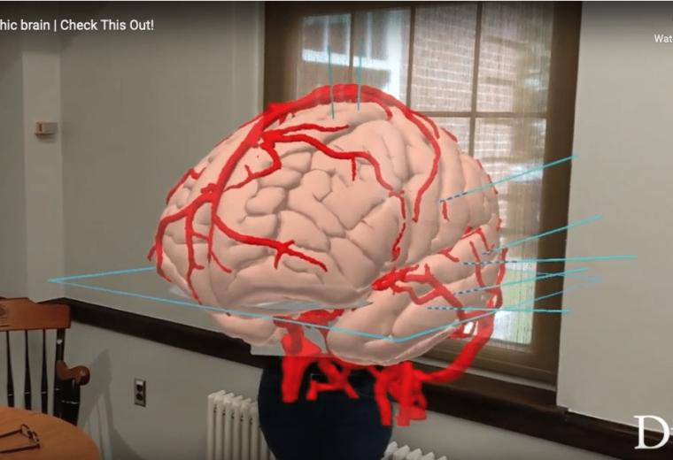
Cameron McIntyre, Ph.D. and colleagues have spent the past seven years developing HoloSNS, a visualization tool that translates human brain scans into interactive holograms

Competitive, five-year grant will help Horstmeyer develop new computational microscopes capable of capturing and processing large-volume, high speed video
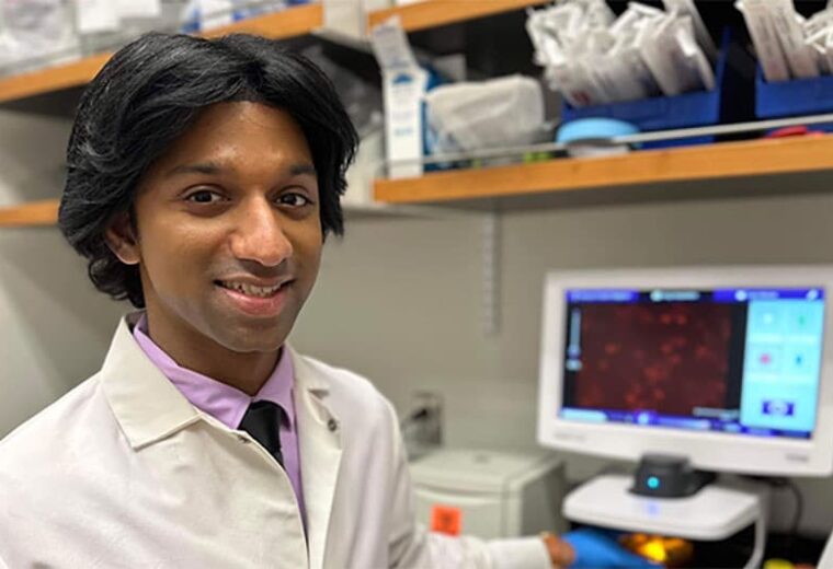
Chatterjee was one of ten recipients of the research award, which will support his project to study a rare form of cancer

New imaging approach visualizes how applying force to proteins alters complex formations
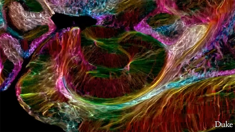
MRI technology from Duke-led effort reveals the entire mouse brain in the highest resolution
Stitching videos from dozens of cameras together provides unique 3D view of macroscopic experiments with microscopic detail
Dalton helps students, staff and faculty learn how to use complex tools to develop miniscule solutions to big problems

The biomedical engineer specializes in using biomaterials to help the body heal damaged tissue, will lead the center with Charles Gersbach