Finding the Signal Through the Noise
By Michaela Kane
Duke BME researchers are pioneering new approaches to turn raw data into valuable insights to detect disease and guide treatment

From imaging technologies that scan many layers of tissue to advanced sensors that can use light to track everything from heart rate to blood sugar levels, researchers are developing tools that can generate biomedical information at an unprecedented scale and pace. Through advancements in artificial intelligence, digital health modeling and data science, Duke BME researchers are pioneering new approaches to turn that raw data into valuable insights to detect disease and guide treatment.
Decoding Data from the Eye
During a standard eye exam, an ophthalmologist may examine the layers of neurons and tissue at the back of the eye. When a person’s eyes are healthy, these layers, each made of different types of cells, appear thick and organized. But if a person has a disease like glaucoma or age-related macular degeneration, these layers may shrink or become deformed.
Researchers can use tools like Optical Coherence Tomography, or OCT, to specifically detect the thickness of these individual layers and determine how far the disease has progressed, which informs the course of treatment. But while these measurements are accurate, they aren’t sensitive enough to detect the early stages of many eye diseases, when individual cells die without affecting the overall thickness and shape of the tissue.
Utilizing the most sensitive imaging system to date, Sina Farsiu, the Paul Ruffin Scarborough Associate Professor of Biomedical Engineering, used his expertise in artificial intelligence to create deep-learning algorithms that can accurately identify subtle biomarkers of eye diseases other than thickness from OCT images of the retina, allowing for earlier diagnosis.
Farsiu is the director of the Vision and Image Processing Laboratory at Duke, where his students pursue projects at the intersection of biomedical engineering, ophthalmology, electrical and computer engineering, and computer science.
“We develop machine learning algorithms for a variety of photonic imaging systems to enhance the quality of images,” says Farsiu. “This work gives us a mountain of data to work with, and we’ve used that data to develop intelligent algorithms that can accurately point out biomarkers of both ophthalmological diseases and neurodegenerative diseases, like Alzheimer’s and ALS.”

These algorithms have also proven useful for clinical trials. For example, a 2020 study showed that researchers could use software built by Farsiu’s PhD student Jessica Loo to accurately assess thousands of images from a phase II clinical drug study.
“This software allowed us to scan the images for biomarkers that indicated whether the drug being tested was working or not,” says Farsiu. “We showed that it was just as accurate as the extremely costly human assessment from a standard clinical trial.”
Now, Farsiu and his postdoc, Reza Rasti, are exploring how they can apply algorithms to predict the outcome of various therapies for patients with diabetes-related vision loss.

The first-line therapy for these patients is typically anti-vascular endothelial growth factor, or anti-VEGF, which can often save a person’s vision for several years.However, the drug must be injected directly into the eye through an invasive and expensive procedure, and it doesn’t work for all patients.“There is a large portion of people where this therapy isn’t effective,” says Farsiu. “If anti-VEGF doesn’t work, then the doctor will laser your eye, and if that doesn’t work, they will explore a series of other treatment options until you respond or you go blind, which isn’t ideal.”With their approach, the team can identify biomarkers from OCT images that indicate which patients would respond to anti-VEGF therapy and which ones won’t, saving them from undergoing an invasive and costly therapy.“This technology is really robust––it has applications for different disease detection and other practical uses,” says Farsiu. “The goal of AI so far is to be able to replicate what doctors do, but no doctor can just look at images and say that a patient will respond to a specific therapy. With this tool, we’ve now gone beyond what a doctor can do, and we can do it with very high accuracy.”

Wearable Devices for a Wealth of Health Insights
Whether it’s on a track, on a trail, in the pool or at the gym, smartwatches and other wearable devices are becoming the most common accessory seen on a person’s wrist. These devices can measure a person’s activity levels, recording an average running pace, daily step counts and even biological data, like heart rate and sleep patterns.
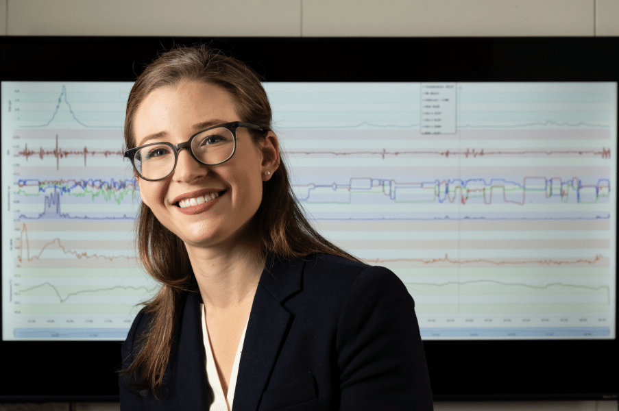
But now, Jessilyn Dunn, an assistant professor of biomedical engineering and biostatistics & bioinformatics at Duke, aims to explore how both researchers and physicians can transform everyday data into effective health decisions.
Dunn runs the Big Ideas Lab, which merges data science and biomedical engineering. With a joint appointment in the department of biostatistics & bioinformatics, Dunn has focused her efforts on understanding how wearable devices can identify digital biomarkers for cardiometabolic diseases, like diabetes, and assessing and improving the accuracy of wearable technology.
“My goal is to turn data sources from an information delivery system into an insight delivery system,” says Dunn. “When you go to the doctor for a check-up, they usually see you for about 15 minutes once a year. Instead, we’d like to use these devices to gather your health data over longer periods and allow physicians to use that data not only to catch who is sick, but to catch who will become sick and then develop appropriate interventions.”
Dunn is exploring this idea through a MEDx (Medicine + Engineering at Duke) grant, which funds a project centered on detecting digital biomarkers of glycemia and glycemic variability. These measurements indicate the amount of glucose in the blood, which determines if a person is prediabetic or diabetic.

“One in three Americans is prediabetic, and that’s detected when a person goes into a clinic for a blood glucose test,” says Dunn. “Ideally, that’s representative of the norm for that person, but if that’s not the case then the diagnosis and subsequent therapies would be missed. We’d like to come up with a more continuous metric that would give more accurate information about someone’s likelihood of becoming prediabetic.”
Building on Dunn’s previous research, the team is developing predictive algorithms and using machine learning to identify digital biomarkers that indicate if a person is at risk of prediabetes.
“This could involve a combination of measuring the number of steps a person takes and their circadian change in heart rate,” says Dunn. “The combination of the change in multiple factors could actually be predictive of someone’s insulin sensitivity, as it gives us an idea of their heart’s function and their physical activity level.”
Allowing patients to gather these measurements via wearable devices removes the need for invasive testing, as patients are much more likely to wear a smartwatch than they are to have regular blood draws.
But as uses for wearable devices continue to grow, Dunn also wants to ensure that these devices are working as accurately as possible for anyone interested in using them.
“Companies that manufacture these devices don’t put out any metrics about how well they work across a variety of factors,” says Dunn. “So we wanted to collect evidence about how well they work and identify potential circumstances where they may not work well.”
In a February 2020 study, Dunn and her PhD student Brinnae Bent explored the accuracy of the heart rate measurements from the most popular wearable devices: the Apple Watch 4, Fitbit Charge 2, Garmin Vivosmart 3 and Xiaomi Midband. The team assessed their accuracy over a variety of skin tones and during various activities, using an ECG measurement as the reference standard.
The team found that, while the devices aren’t affected by skin tones, the accuracy between wearables begins to vary wildly when they measure heart rate during different types of everyday activities, such as typing or walking.
“If there are disparities in how these devices work, we need to identify and correct them,” says Dunn. “Our hope is that these wearables will fill a gap in the health care world. Health happens 24 hours a day, seven days a week. Our goal is to improve long-term and acute health monitoring outside of the clinic and get easy-to-use health care tools to people who may not typically have easy access to standard health care in the first place.”
